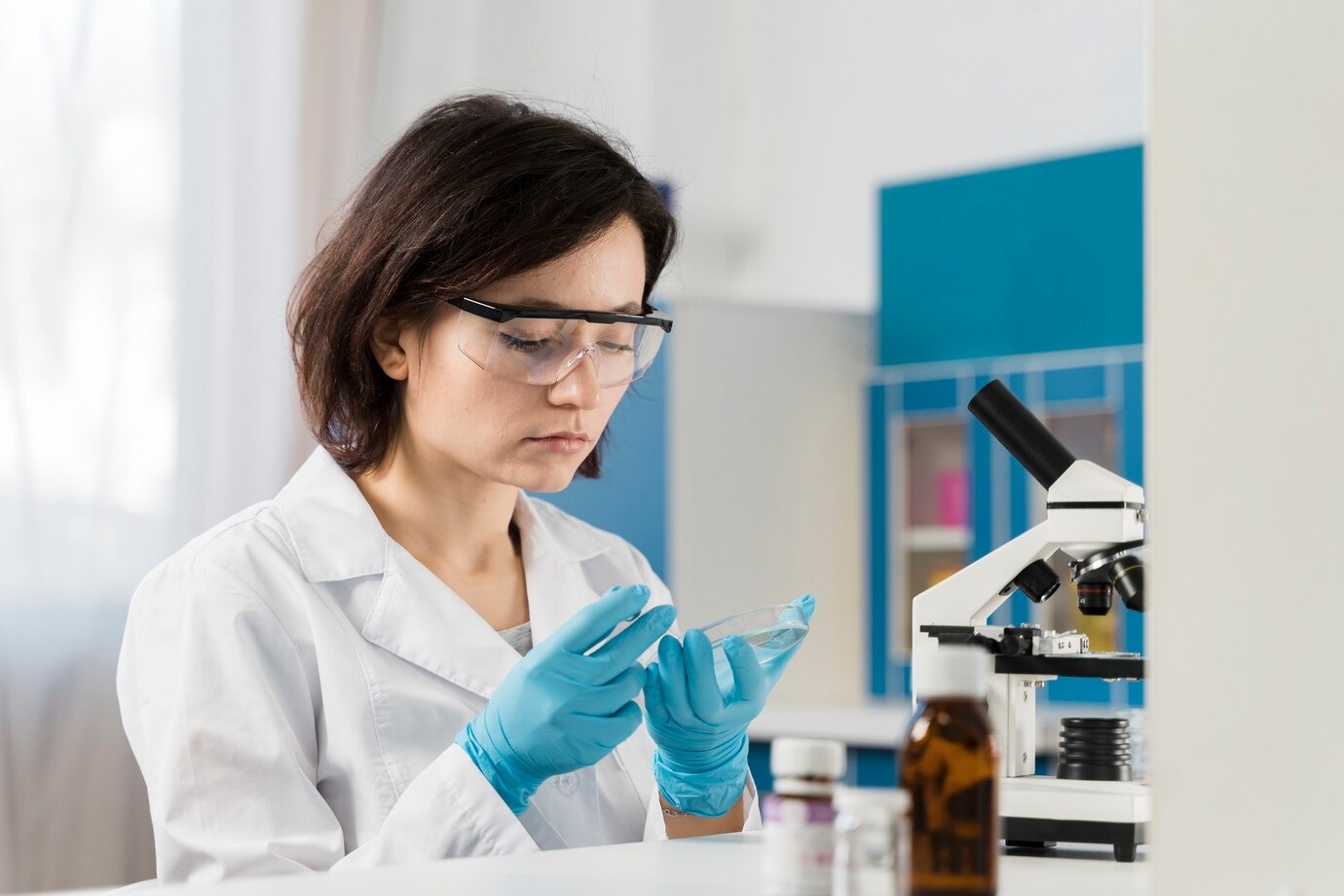
I still remember the first time I performed SDS PAGE electrophoresis in my lab. The glass plates, the gels, the buffer solutions—it all felt like a delicate art form. Over the years, I’ve refined my technique, learned from mistakes, and developed a step-by-step process that consistently delivers accurate and reproducible protein separation. If you’re aiming to master SDS PAGE in your lab, here’s the approach that has worked for me, from preparation to analysis.
Step 1: Understand the Basics Before You Begin
Before even touching the equipment, I made sure I understood the core principle of SDS PAGE. This technique separates proteins based on molecular weight. SDS (sodium dodecyl sulfate) denatures proteins and coats them with a negative charge, allowing them to migrate through the gel according to size when an electric current is applied.
Knowing why you are doing each step ensures you make informed decisions during the process. Without this foundation, mistakes are harder to identify and correct.
Step 2: Prepare Your Samples Properly
I learned early on that sample preparation is the most critical step for clear, distinct protein bands. My process includes:
- Protein Quantification: Before loading samples, I measure protein concentration using a Bradford or BCA assay. This ensures equal loading and accurate comparison.
- Adding Loading Buffer: I mix my protein with SDS loading buffer containing β-mercaptoethanol or DTT. This breaks disulfide bonds and ensures complete denaturation.
- Heating Samples: I incubate them at 95°C for 5 minutes (some sensitive proteins may require less heat).
Poor preparation at this stage often leads to smeared bands or uneven migration.
Step 3: Select the Right Gel Percentage
One mistake I made in my early experiments was using the wrong gel concentration for my target proteins. Now, I always choose gel percentage based on the molecular weight range:
- 4–8% gel for large proteins (>200 kDa)
- 10–12% gel for medium proteins (30–200 kDa)
- 15% gel for small proteins (<30 kDa)
Sometimes, I prepare a gradient gel if my sample contains a wide size range. This small adjustment often makes the difference between a clean result and a frustrating repeat experiment.
Step 4: Cast the Gel Without Bubbles
When pouring the resolving gel, I pay special attention to avoid bubbles. Bubbles can distort protein migration and ruin the run. I also overlay the gel with a thin layer of isopropanol to create a flat surface and prevent oxygen inhibition.
Once polymerized, I remove the alcohol, rinse with distilled water, and carefully pour the stacking gel on top. The stacking gel ensures proteins start migrating in a tight band before entering the resolving gel.
Step 5: Assemble the Apparatus Correctly
Misaligning gel plates or forgetting to fill the inner chamber with buffer was something I learned to avoid the hard way. I always double-check that:
- Plates are firmly clamped without leaks.
- The comb is removed without damaging wells.
- The running buffer completely covers the wells and electrodes.
A poorly assembled apparatus leads to uneven runs and wasted samples.
Step 6: Load Samples and Marker with Steady Hands
Loading samples into the wells requires precision. I use a micropipette and make sure to slowly dispense the sample at the bottom of the well without piercing it. A pre-stained protein marker is always loaded in one lane to monitor progress and molecular weight.
Consistency in loading volume and concentration ensures reliable band comparison across lanes.
Step 7: Run the Gel at the Right Voltage
From my experience, rushing the process with high voltage causes overheating and distorted bands. I start the run at 80V during stacking, then increase to 120–150V for the resolving gel.
I also keep an eye on the migration front. Once the dye front is near the bottom, I stop the run to avoid proteins running off the gel.
Step 8: Stain and Destain for Clear Visualization
If I’m doing Coomassie staining, I immerse the gel in stain for about an hour, then destain overnight. For more sensitivity, I use silver staining, which can detect nanogram levels of protein.
For Western blotting, I skip full staining and transfer proteins directly to a membrane for antibody detection. Choosing the right visualization method depends on the purpose of the experiment.
Step 9: Analyze Your Results
I take photographs of the gel under consistent lighting conditions. Then, I measure band intensity and migration distance using image analysis software.
If I notice faint or smeared bands, I revisit my sample preparation or gel percentage. Analysis isn’t just about recording results—it’s about identifying areas for improvement in the next run.
Step 10: Troubleshoot Like a Scientist
Even after years of practice, SDS PAGE sometimes throws surprises. Here’s my personal troubleshooting checklist:
- Smeared bands: Could be due to overloaded wells, degraded samples, or high salt concentration.
- No bands: Possible issues include incorrect sample preparation, broken circuit, or expired reagents.
- Uneven migration: Likely from poor gel polymerization or uneven electrode contact.
Rather than feeling frustrated, I treat these issues as learning opportunities.
Step 11: Maintain Your Equipment
I’ve learned that well-maintained equipment saves time and ensures reproducibility. After each run, I rinse gel plates, combs, and chambers with distilled water and mild detergent. I also regularly check for cracks, warping, or rust in clamps and electrodes.
Neglecting equipment care often shows up as inconsistent results.
Step 12: Keep a Detailed Lab Notebook
Every time I run SDS PAGE, I document:
- Gel percentage and composition
- Sample type and preparation steps
- Voltage, run time, and temperature
- Staining method and results
This record-keeping has saved me countless hours when I need to replicate a successful experiment or troubleshoot an unexpected outcome.
My Final Advice for Mastering SDS PAGE
Mastering SDS PAGE isn’t about rushing through the steps—it’s about paying attention to detail and understanding why each step matters. When I first started, I focused only on the physical process. Over time, I learned that success depends equally on preparation, precision, and post-run analysis.
Once you grasp the technique, you’ll find SDS PAGE to be one of the most powerful tools for protein analysis in your lab. It will help you uncover protein expression patterns, verify purification results, and advance your research with confidence.
Visit our website for more resources and expert guidance on mastering SDS PAGE electrophoresis.
Contact Us


